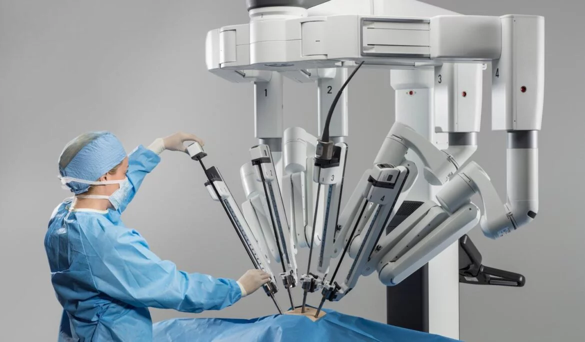
Robotic-Assisted Nephroureterectomy
(Minimally Invasive Kidney and Ureter Removal Surgery)
Robotic-assisted nephro-ureterectomy is a minimally invasive surgical procedure used to treat cancers arising from the inner lining of the kidney or ureter, known as urothelial tumours. These cancers are similar to bladder cancer and can occur anywhere along the lining of the urinary tract.
Using advanced robotic technology, the entire kidney and ureter are removed through a keyhole approach. This is essential to reduce the risk of cancer recurrence, as urothelial tumours have a tendency to recur if any affected tissue is left behind.
At Urology SA, Dr Jimmy Lam is highly skilled in performing this procedure using the da Vinci Surgical System—a robotic-assisted platform designed to enhance surgical precision and patient outcomes.
Who is suitable for robotic-assisted nephro-ureterectomy?
This procedure may be recommended for patients diagnosed with:
- Urothelial cancer of the renal pelvis (the central part of the kidney that collects urine)
- Urothelial tumours of the ureter (the tube connecting the kidney to the bladder)
Your surgeon will assess your individual condition, imaging results, and overall health to determine whether this approach is suitable for you.

Benefits of robotic-assisted nephro-ureterectomy
Compared to traditional open or standard laparoscopic surgery, robotic-assisted nephro-ureterectomy offers several key advantages:
For the patient:
- Smaller incisions and minimal scarring
- Reduced blood loss during surgery
- Shorter hospital stay and quicker recovery
- No need for large incisions or cuts into the bladder
For the surgeon:
- High-definition, 3D magnified vision
- Robotic arms with a greater range of motion than the human hand
- Improved dexterity and precision, especially in delicate areas such as where the ureter meets the bladder
How the procedure is performed
You will be under general anaesthetic for this procedure, which typically takes around 3 hours.
The steps include:
- 3–5 small keyhole incisions are made in the abdomen.
- A camera and robotic surgical instruments are inserted.
- The abdomen is inflated with carbon dioxide gas to create space and improve visibility.
- The entire kidney and ureter are carefully removed under robotic guidance.
- The ureter is detached from the bladder, and the small opening in the bladder is meticulously closed to ensure it is water-tight.
- The kidney and ureter are removed from the body through one of the incisions.
- The gas is released, instruments are removed, and the incisions are closed carefully to minimise scarring and reduce the risk of hernia.
What to expect after surgery
- Most patients stay in hospital for 2–3 nights.
- Mild to moderate pain or discomfort is normal and can be effectively managed with medications.
- You will be encouraged to:
- Sit out of bed and walk early
- Perform deep-breathing exercises to reduce the risk of pneumonia or blood clots (DVT/PE)
Uncommon complications may include:
- Urine leakage from the bladder repair site
- Injury to major blood vessels or nearby organs
- Incisional hernia, especially if wound healing is impaired
Your care team will provide specific post-operative instructions and monitor your recovery closely. If you have any concerns after discharge, contact the clinic or, if urgent, attend your nearest emergency department.
